Sunday, October 30, 2011

Specimen #9 Cup Fungi


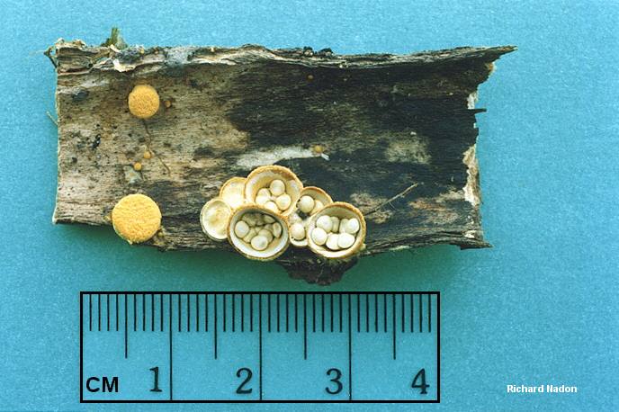
Fig. 1 Eyelash Cup Fungus showing the miniscule
 hairs along the exterior and margin of the fungus
 (Stevens & Woods,
2009). 
Name: Scutellinia scutellata -Eyelash Pixie Cup
Family: Pyronemataceae
Collection Date: November 8, 2011
Habitat: Growing on wood
Location: Hiram Field Station
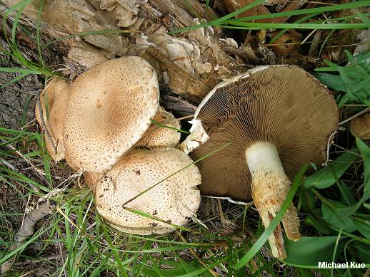
Description: "At first spherical, but soon opens to form a shallow cup and eventually a disklike (flattened). Fertile (upper) surface smooth, bright red to scarlet to orange (or rarely paler with a pinkish cast); margin conspiciously ciliate (fringed with dark brown or blackish hairs up to 1 mm long). Exterior (underside) also clothed with dark hairs. Flesh very thin. Stalk absent. Found growing on rotten wood or damp soil ( or occasionally on ashes, wet leaves, or conks); widely distributed and common, but easily overlooked because of its small size" (Aurora, 1986).
"Spores... smooth when immature, and remaining so for a long time--but in maturity prominently sculpted with warts and ribs extending to about 1 µ high; with several oil droplets. Paraphyses with swollen tips 6-10 µ across" (Kuo, 2009).
Collector: Brooke Warren
Key Used: Aurora, D. (1986). Mushrooms Demystified. New York: Ten Speed Press.
Keying Steps:
Key to the Major Groups of Fleshy Fungi p. 52
1B. Spores produced inside mother cells called asci; fruiting body variously shaped (see pg. 55).... Ascomycotina, p. 782

Key to the Ascomycetes p. 782
1B. Not as above; growing on wood or on ground, on insects, other mushrooms, plants, etc... 2
2A. Growing on wood (but wood sometimes buried)... 3
3B. Fruiting body cuplike or variously shaped but not as above, or if colored as above then texture usually different (fragile, fleshy, rubbery, gelatinous, etc.); asci typically borne in a palisade (hymenium), not in perithecia... Discomycetes, p. 783
Key to Discomycetes
1B. Not as above; fruiting body occasionally buried but usually above the ground at muturity or on wood, moss, etc.; spore-bearing surface exposed (external) at maturity... 2
2A. Fruiting body cup- to ear-shaped, disklike (flat), cushion-like, top-shaped, or sometimes contorted; stalk absent or present only as a narrowed base (but fruiting body sometimes growing erect like a rabbit's ear); asci operculate (i.e., with "lid" at tip)... Pezizales p.783

Key to Pezizales
1B. Not as above... 3
3A. Fruiting body cup-shaped (concave) to disk-like (flat), cushion-shaped, or sometimes top-shaped or splitting into rays; stalk present or absent; flesh gelatinous, fragile, or tough... 4
4A. Stalk absent, or if present then often (but not always) short or merely a narrowed, downward extension of the cup; stalk when present usually lacking distinct ribs; fruiting body fleshy, fragile, rubbery, or gelatinous, sometimes brightly colored, large to minute; tips of asci amyloid or not amyloid.... Pezizacea & Allies, p. 817

Key to the Pezizacea & Allies
1B. Not as above... 2
2B. Not with above features (but may have some of them)... 3
3B. Not as above... 4
4B. Not as above; fruiting body cuplike, earlike, disclike, etc. (but sometimes contorted, especially if growing in clusters)... 6
6B. Not as above; fruiting bodies sometims slit down one side but not consistently so, and not usually growing erect; sometimes growing on dung... 7
7A. Fertile (upper or inner) surface of fruiting body brightly colored (red, orange, yellow, blue, or green, but not violet)... Aleuria & Allies, p. 833

Key to Aleuria & Allies
1B. Not as above... 3
3A. Exterior (underside) of cup or disc clothed with brown to black hairs; margin often fringed with dark hairs also... 4
4B. Not as above... 5
5A. Fertile (upper) surface yellow, orange, or red... 6
6B. Not as above; growing in soil, humus, or on wood but not usually in burned areas... 7
7B. Not as above... 8
8B. Underside of fruiting body with fairly obvious hairs which fringe the margin like eyelashes; fertile surface bright red to orange-red or orange... Scutellina scutellata & others, p. 839

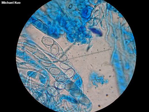
Fig. 2 The spores of this fungus are so small they are unable to be seen
with the naked eye. Here a microscope enables us to see what they look like (Kuo, 2009).
Scutellinia scutellata
Fig. 3 Eyelash Cup Fungi again showing the hairs on the exterior
and margins of the fungus (Wolf, 2011).
Fig. 3 Eyelash Fungi growing on a decaying tree (Brooke Warren).


Additional References:
Kuo, M. (2009, April). The eyelash cup: Scutellinia scutellata. Retrieved from the MushroomExpert.Com Web site: http://www.mushroomexpert.com/scutellinia_scutellata.html
Stevens, F. & Woods, M. (2009). California Fungi- Scutellina scutella. The Fungi of California. http://www.mykoweb.com/CAF/species/Scutellinia_scutellata.html
Wolf, R. (2011). Scutellinia scutellata. Calphotos. http://calphotos.berkeley.edu/cgi/img_query?stat=BROWSE&query_src=photos_fungi_com&where-genre=Fungi&where-namesoup=Eyelash+Pixie+Cup&rel-namesoup=matchphrase&title_tag=Eyelash+Pixie+Cup


Links:
http://www.mushroomexpert.com/scutellinia_scutellata.html
http://www.mykoweb.com/CAF/species/Scutellinia_scutellata.html

Specimen #8 Agarics (gilled) Fungus

Name: Mycena murina  Family: Mycenaceae
Collection date: October 8, 2011
Habitat: Growing in a cluster under a pine
Location: Hiram, ohio
Description: The pileus can be up to 15 mm across, conical to convex, Sometimes with a point in the middle that eventually flattens with age. Striated, lubricous to viscid, darker in the center and becoming paler grey or grey-brown towards the margin. The stipe is hollow and can be straight up or flexuous, glutinous or viscid (Aronsen, 2011).
Collector: Brooke Warren
KeyUsed:Arora, D. (1986). Mushrooms Demystified.New York: Ten Speed Press.
Keying steps:
Key to the Major Groups of Fleshy Fungi
1A. Spores produced on mother cells called basidia; fruiting body variously shaped... Basidiomycotina p. 57

Key to the Basidiomycetes1A. Basidia and spores borne externally (on the exposed surfaces of gills, tubes, spines,
 branches, lobes, etc.); spores forcibly discharged at maturity, i.e., a spore print often (but not always) obtainable;
fruiting body with a cap and stalk, or clublike, orbranched, or bracketlike, or crustlike (without a stalk or sometimes without a cap) or lobed  or bloblike, etc. ... 2
2B. Not as above... Hymenomycetes p. 58


Key to the Hymenomycetes1B. Not as above; pores and tubes absent... 3
3A. Underside of cap with radiating blades (gills)... Agaricales p. 59


Key to the Agaricales
1B. Not as above; spores forcibly discharged, hence a spore print obtainable if spores are being produced; gills
exposed at maturity; common and widespread... 2
2A. Spore print white to buff, yellow, yellow-orange, or lilac-tinged...3
3B. Neither volva nor warts present (but cap and stalk may have scales or fibrils)... 4
4B. Not as above; veil absent, or if present then gills normally attached to stalk... 6
6B. Not as above; gills usually platelike or bladelike... 7
7B. Not as above... 8
8B. Not with above features... 9
9B. Not as above; gills not normally waxy; stalk central to lateral or absent; on ground or wood...
Tricholomataceae, p. 129

Key to Tricholomateae
1B. Not growing on other mushrooms, or if so then gills well-developed, thin, close... 2
2B. Not as above... 3
3B. Stalk present, well-developed, more or less central; growing on ground or wood... 6
6B. Not as above (but stalk may have a tapered underground "tap root")... 7
7B. Not as above; weil absent, or if present then cap and stalk not granulose... 8
8B. Veil absent or rudimentary and evanescent, not forming an annulus... 9
9B. Not as above... 10
10B. Stalk usually thin and hollow or stuffed and either fragile or cartilaginous (tough), typically 5 mm
thick or less (occassionally thicker but then with a tough cartilaginous outer rind)... 23
23B. Not as above... 24
24A. Cap conical or bell-shaped when young (but may expand in age), often translucent-striate when moist,
margin not usually incurved when young; stalk not polished or tough... Mycena, p. 224


Additional References:
Aronsen, A. (2003). A Key to the Mycenas of Norway. Mycena Page. http://www.bing.com/images/search?q=mycena+vulgaris&view=detail&id=8352DFF86E54B73351BC3E7DF139D3680D52AD9B&first=
0&qpvt=mycena+vulgaris&FORM=IDFRIR

Links:
http://www.mycokey.com/MycoKeySolidState/species/Mycena_vulgaris.html
http://www.bing.com/images/search?q=mycena+vulgaris&view=detail&id=8352DFF86E54B73351BC3E7DF139D3680D52AD9B&first=
0&qpvt=mycena+vulgaris&FORM=IDFRIR
Mycena vulgaris , klæbrig huesvamp, klisterhette, klibbhætta, Kleefsnedemycena, Klebriger Helmling
Fig. 2 Lamellae with gelatinized edge Illustration and © Jens H. Petersen

Fig. 1 The cap (Horna Fure, n.d.).


Sunday, October 23, 2011

Specimen #7 Bird's Nest Fungus

Fig.1 Cluster of Bird's Nest Fungus (Barron, n.d.).
Name: Crucibulum laeve -Common Bird's Nest Fungus 
Family: Nidulariaceae
Collection Date: October 8, 2011
Habitat: Growing in mulch
Location: Outside of Hiram College's Gerstacker building in Hiram, Ohio
Description: "The fruiting body is tiny, at first nearly round, becoming cylindrical and then deeply cup-shaped; 5-12 mm high and broad at the top when mature, the rim is more or less circular and covered at first by a hairy lid. Peridium (wall of nest) is one-layered, tough, and persistent. Exterior is vely or shaggy, yellowish or tawny to cinnamon-brown, becoming nearly smooth in age and often darker or whiter. Interior of nest is smooth and somewhat shiny, white, to silvery, gray, or pale cinnamon. The peridioles (eggs) 1-2 mm in diameter, several, whitish to buff or with a very slight brownish tinge, circular but flattened (lens- or disclike), usually attached to nest by long thincords. Widely distributed. Scattered to densely gregarious on sticks, wood chips, nut shells, vegetable debris, humus, and manure" (Arora p. 779, 1986).
Collector: Brooke Warren
Key Used: Arora, D. (1986). Mushrooms Demystified. New York: Ten Speed Press.
Keying Steps:
Key to the Major Groups of Fleshy Fungi
1A. Spores produced on mother cells called basidia; fruiting body variously shaped (see pp. 52-54)... Basidiomycotina, p. 57

Key to the Basidiomycetes
1B. Basidia and spores borne internally (inside the fruiting body or inside a spore case or small capsules); spores not forcibly discharged; thus a spore print is unobtainable... Gasteromycetes, p. 676

Key to the Gasteromycetes
1A. Fruiting body minute (typically less than 15 mm high), consisting of a "nest" (cup, vase, or bowl) containing one or more "eggs" (periodioles); (older specimens, however, may lack peridioles and young ones often have a covering or "lid" pver the top of the "nest")... Nidulariales, p. 778

Key to the Nidulariales
1B. Not as above; fruiting body cylindrical to mug- or cup- shaped when mature, containing more than one "egg" (unless all but one has been expelled)... 2
2B. Not as above; fruiting body typically with a "lid" when very young, the nest usually well formed and persistent; eggs may or may not be imbedded in a mucilage... 4
4B. Eggs white to gray, brown, or black, often (but not always) attached to the side of the nest by a minute cord or short stalk, not imbedded in a mucilage; sides of nest vertical to tapered... 6
6B. Interior of nest smooth or at least not striate or grooved... 7
7A. Eggs typically white to buff; interior of nest not black... Crucibulum laeve (& others)


Fig. 2 Growing on mulch. Easy to see why it is called the Bird's nest fungus.
The Peridium resembles a nest while the Peridioles resemble eggs inside the nest


Crucibulum laeve
Fig.1 Example of the actual size of this fungus (Kuo, 2003).


Fig. 3 Side view of the peridium
Fig. 4 View of the fungus as if you were walking by it. As
you can see it isgrowing in the mulch in clusters
Fig.5 Another example of how small this fungus truly is. If you look closely
you can see how capsule hasn't been lost yet (Kuo, 2003).


Links:
http://www.mushroomexpert.com/crucibulum_laeve.html
http://www.rogersmushrooms.com/gallery/DisplayBlock~bid~5881.asp

Additonal Citations:
Kuo, M. (2003, September). Crucibulum laeve. Retrieved from the MushroomExpert.Com Web site: http://www.mushroomexpert.com/crucibulum_laeve.html
Barron, G. (n.d.). Crucibulum laeve. Retrieved from George Barron's Website on Fungi Website: http://www.uoguelph.ca/~gbarron/GASTEROS/crucibul.htm

Specimen #6 Puffball Fungus

Name: Sleroderma citrinum -Common Earthball
Family: Sclerodermataceae
Collection Date: September 27,2011
Habitat: Growing under conifer's in a small group and also found on a decaying tree in another area
Location: James H . Barrow Field Station in Hiram, Ohio
Description: "The fruiting body is about 2-10 cm broad and about 2-6 cm high, round or somewhat flattened, the underside often with a stemlike base composed of ridges, mycelial fibers, and/or adhering debris; evntually cracking into lobes to form an irregular pore, the lobes not normally bending outward or unfolding appreciably. The peridium is hard and fairly rigid, 1-3 mm thick, white when sectioned, usually staining pinkish if fresh; the surface is yellow-brown to tan and cracked or arranged into prominent, inherent scales which often have a smaller, central wart. The spore mass is white when very young, soon gray to purple-gray with whitish veins running through it, then drak purple-gray to purple-black or black and still solid and firm; eventually becoming powdery and blackish-brown. They can be solitary, scattered, or in groups or clumps on the ground or in rotten wood. Very widespread and common. They are poisonous! Can cause nausea, vomiting, diarrhea, chills, and cold sweats" (Aurora p. 708, 1986).
Collector: Brooke Warren
Key Used: Aurora, D. (1986). Mushrooms Demystified. New York: Ten Speed Press.
Keying Steps:
Key to the Major Groups of Fleshy Fungi
1A. Spores produced on mother cells called basidia; fruiting body variously shaped (see pp. 52-54)... Basidiomycotina, p. 57

Key to the Basidiomycetes
1B. Basidia and spores borne internally (inside the fruiting body or inside a spore case or small capsules); spores not forcibly discharged, thus a spore print unobtainable Gasteromycetes, p. 676

Key to the Gasteromycetes
1B. Fruiting body differently constructed and usually larger than above... 2
2B. Not as above (but fruting body may be slimy or malodorous at some stage)... 3
3B. Stalk absent or rudimentary... 7
7A. spore case rupturing or disinteggrating at maturity; spore mass firm and solid when young (chambers present hardly discernible), powdery or cottony when mature and usually dispersing fairly soon; columella (internal stalk) typically absent; mature fruiting body usually (but not always) above ground; found in many habitats... Lycoperdales & Allies, p.677

Key to the Lycoperdales & Allies
1B. Not as above (fruiting body may rupture in starlike fashion, but if so then there is no separate spore case within)... 2
2B. Spore mass not containing peridioles, or if so then the peridioles considerably larger than grains of sand (usually aappearing more like seeds)... 4
4B. Not as above; peridioles absent; spores produced in a single large chamber (the spore case)... 5
5A. Spore case typically hard or tough with a thick rindlike skin, at least when young; spore mass white when very young but soon darkening (usually purpole-gray to black) while remaining firm, eventually becoming dark brown to blackish and powdery; basidia not borne in a hymenium; capillitium absent... Scleroderma, p. 707

Key to Scleroderma
1B. Not as above; mature spores ornamented; founnd underground or above; common... 2
2B. Not as above; fruiting above the ground, or if underground then usually with an obvious base or point of attachment; peridium not marbled in cross-section; spores borne on basidia... 3
3B. Not as above; either peridium thinner (averaging 1-4 mm) or not rupturing into starlike lobes; fruiting body fairly small to medium-sized (rarely large)... 4
4B. Not as above ("stalk" if present shorter and/or habitat different)... 5
5A. Peridium (skin) covered with prominent inherent rosette-like scales (i.e. each scale often with a central wart); widespread, but especially common in forests... 6
6B. Peridium fairly thick (usually 1-4 mm), rupturing into lobes in age or forming an irregular pore; spores reticulate... S. citrinum


Fig.1 Group of fruiting bodies that were found growing together under a tree.

Fig. Stem-like base composed of mycelial fibers and debris


Fig. Inside view of the mature purple-gray spore mass
Fig. Yellow-brown colored fruiting body. The scales are a dark brown color


Links:
http://www.norfolkwildlifetrust.org.uk/Wildlife-in-Norfolk/Species/Fungi/Common-Earthball.aspx
http://www.wsbrc.org.uk/uf6YqGEw8EW1y9cezXBvsw%3D%3D/Species.aspx
http://www.first-nature.com/fungi/scleroderma-citrinum.php
http://healing-mushrooms.net/archives/scleroderma-citrinum.html

Tuesday, October 11, 2011

Speciman #1 Flask Fungus


Fig. 1 Cluster of Dead Man's Fingers
Name: Xylaria polymorpha -Dead Man's Fingers
Family: Xylariaceae
Collection Date: October 4, 2011
Habitat: Growing in a cluster of about 6, on a sandy slope near a decaying tree.
Location: The West Woods in Russell, Ohio
Description: "The fruiting body is around 2-8 cm tall, 0.5-3 cm thick, very tough and hard, erect, club or finger shaped, and twisted. The outer surface is hard and crustlike, usually wrinkled, roughened, and / or cracked, black when mature but often covered with a whitish to grayish or brownish powder when very young. The interior flesh is hard or corky, white, or pallid" (Arora, 1986).
Collector: Brooke Warren
Keys Used: Aurora, D. (1986). Mushrooms Demystified. New York: Ten Speed Press.
Keying Steps:
Key to the Major Groups of Fleshy Fungi p. 52
1B. Spores produced inside mother cells called asci; fruiting body variously shaped (see pg. 55).... Ascomycotina, p. 782

Key to the Ascomycetes p. 782
1B. Not as above; growing on wood or on ground, on insects, other mushrooms, plants, etc... 2
2A. Growing on wood (but wood sometimes buried)... 3
3A. Fruiting body usually black or very dark brown (but often covered with white or grayish powder), rounded to irregularly knobby and charcoal-like or fingerlike to clublike or antlerlike and very tough or hard; asci borne in flasklike nests (perithecia) which often give the fertile area of fruiting body a minutely pimpled appearance... Pyrenomycetes, p. 878

Key to the Pyrenomycetes p. 878
1A. Growing on wood (but wood sometimes buried)... 2
2B. Fruiting body gray to dark brown or black, but sometimes covered with a white powder; common... Xylaria & Daldinia, p. 885

Key to Xylaria & Daldinia
1B. Not as above; fruiting body erect, clublike (unbranched) or antlerlike (branched)... 2
2B. Not as above... 3
3A. Fruiting body very tough or hard, up to 3 cm thick; flesh inside usually white or pallid; surface often minutely warted or cracked... Xylaria polymorpha & others, p. 886
Fig. 2 Inside flesh (white part) of a dead man's finger
fruiting body
Fig. 3 An example of how tall a fruiting body
of a Dead Man's Finger is


Links
http://www.mushroomexpert.com/xylaria_polymorpha.html
http://botit.botany.wisc.edu/toms_fungi/apr2000.html
http://www.psu.edu/dept/nkbiology/naturetrail/speciespages/deadmansfingers.htm
http://www.messiah.edu/Oakes/fungi_on_wood/club%20and%20coral/species%20pages/Xylaria%20polymorpha.htm

Specimen #4 Agaric (Gilled) Fungus


Fig. 1 Cluster of Shaggy Mane fungi. The cap appears shaggy
due to the remnants of the universal veil still attached (Kuo, 2008).
Name: Coprinus comatus -Shaggy Mane
Family: Agaricaceae
Collection date: October 5, 2011
Habitat: Growing individually along the side walk in grass and mulch.
Location: Along the side walk that is across from the Hiram Post Office and next to the pizza shop building. Hiram, Ohio

Description: "The cap is about 4-15 cm tall, cylindrical or columnar, expanding somewhat as margin curls up until it is more or less bell-shaped, then deliquescing from the bottom up; surface not viscid, white with brown to pale cinnamon-brown or buff center, soon breaking up into shaggy white to brown scales (universal veil remnants) which often recurve in age; margin striate in age and often tattered. Flesh soft, white. The gills are very crowded. At first white and then pass through pink shades until it finally becomes black and inky" (Arora p. 345, 1986).
Collector: Brooke Warren
Key Used: Arora, D. (1986). Mushrooms Demystified. New York: Ten Speed Press.
Keying Steps:
Key to the Major Groups of Fleshy Fungi
1A. Spores produced on mother cells called basidia; fruiting body variously shaped (see pp. 52-54)... Basidiomycotina, p. 57

Key to the Agaricales

1B. Not as above; spores forcibly discharged, hence a spore print obtainable if spores are being produced; gills exposed at maturity; common and widespread... 2
2B. Spore print some other color (pinkish, salmon, yellow-brown, brown, rusty-orange, rusty- brown, chocolate-brown, purplish, greenish, black, etc.)... 10
10B. Spore print some shade of orange, brown (including cinnamon-brown), green, purple, gray, or black...16
16B. Not as above... 19
19A. Spore print purple-brown to purple-gray, purple-black, smoky-gray, black, chocolate-brown, or deep brown... 20
20B. Not as above; gills free to adnexed, adnate, or occasionally decurrent... 21
21A. Gills and/ or cap auto-digesting (i.e., turning into an inky black mass) at maturity; spore print black... Coprinaceae, P. 341

Key to the Coprinaceae
1A. Mature gills (and often the cap) digesting themselves, i.e., either turning into an inky black fluid or withering away... Coprinus, p. 342

Key to Coprinus
1B. Growing on ground, wood chips, wood, or indoors... 9
9A. Cap cylindrical before expansion and4-25 cm or more tall, usually at least somewhat shaggy, entirely white or with a brown center and / or brownish scales; partial veil present when young, often forming a movable annulus (ring) that may drop off; cosmopolitan.... C. comatus, p. 345

"Their mass rotted off them flake by flake
Til the thick stalk stuck like a murderer;s stake,
Where rags of loose flesh yet tremble on high
Infecting the winds that wander by" (Arora p.342, 1986).


Coprinus comatus
Fig. 2 Deliquescingfrom the bottom up. The Shaggy Mane's way of
releasing its spores into the environment (Kuo, 2008).

Coprinus comatus
Fig. 3 This fungus's fruiting body comes in many different sizes (Kuo, 2008).
Fig. 4 Shows the stem of the Shaggy Mane. As the picture shows it is easily
separable from the cap; hollow (Kuo, 2008).

Fig. 5 Shaggy Mane found in Hiram, Ohio. Looks like an egg sitting in the
 grass when the stem isn't visible.


Links:
http://www.mushroomexpert.com/coprinus_comatus.html
 http://botit.botany.wisc.edu/toms_fungi/may2004.html
http://healing-mushrooms.net/archives/9.html
http://youtu.be/Jth5IMq44Ec

Citations:
Kuo, M. (2008, February). Coprinus comatus: The shaggy mane. Retrieved from the MushroomExpert.Com Web site: http://www.mushroomexpert.com/coprinus_comatus.html

Specimen #5 Agaric (Gilled) Fungus

Fig.1 Cluster of P. squarrosa on a decaying log


Name: Pholiota squarrosa -Scaly Pholiota
Family: Strophariaceae
Collection Date: October 4, 2011
Habitat: Growing on dead tree
Location: The West Woods in Russell, Ohio
Description: "The cap is 3-10 cm  broad, obtuse or convex becoming broadly bell-shaped to slightly
umbonate or plane; surface dry, pale tan to straw colored, buff, or pale yellow-brown, or in age darker
yellow-brown or sometimes greenish-yellow toward the margin; covered with a dense layer of upright or recurved, often darker brown) scales; margin incurved at first and often fringed copiously with veil remnants. Flesh pale yellowish. Odor is mild; garlic or onion-like. The gills are crowded" (Arora p. 390, 1986).
Collector: Brooke Warren
Key Used: Arora, D. (1986). Mushrooms Demystified. New York: Ten Speed Press.
Keying Steps:
Key to the Major Groups of Fleshy Fungi
1A. Spores produced on mother cells called basidia; fruiting body variously shaped (see pp. 52-54)...
Basidiomycotina, p. 57

Key to the Basidiomycetes
1A. Basidia and spores borne externally (on the exposed surfaces of gills, tubes, spines, branches, lobes, etc.); spores forcibly discharged at
maturity, i.e., a spore print often (but not always) obtainable; fruiting body with a cap and stalk, or clublike, or branched, or bracketlike, or
crustlike (without a stalk or sometimes without a cap) or lobed  or bloblike, etc. ... 2
2B. Not as above... Hymenomycetes, below

Key to the Hymenomycetes
1B. Not as above; pores and tubes absent... 3
3A. Underside of cap with radiating blades (gills)... Agaricales, below

Key to the Agaricales
1B. Not as above; spores forcibly discharged, hence a spore print obtainable if spores are being produced; gills exposed at maturity; common
and widespread... 2
2B. Spore print some other color (pinkish, salmon, yellow-brown, brown, rusty-orange, rusty brown, chocolate-brown, purplish, greenish, black,
etc.)... 10
10B. Spore print some shade of orange, brown, (including cinnamon-brown), green, purple, gray, or black... 16
16B. Not as above... 19
19A. Spore print purple-brown to purple-gray, purple-black, smoky-gray, black, chocolate-brown, or deep brown... 20
20B. Not as above; gills free to adnexed, adnate, or occasionally decurrent... 21
21B. Not as above... 22
22B. Not as above... 23
23B. Cap dull (some shade of brown, buff, gray, white, etc.)... 24
24B. Not as above... 25
25A. Cap usually viscid when moist (but may dry out!); fruiting body fragile or not fragile; cap cuticle typically filamentous (under microscope)...
Strophariaceae, p. 367

Key to the Strophariaceae
1A. Spore print dull to brown to cinnamon-brown or rusty-brown... Pholiota, p. 384

Key to Pholiota
1B. Not typically growing in ashes... 3
3B. Not as above ( if blue or greenish-blue when young then found in eastern North America)... 4
4A. Growing on wood (occasionally buried) or in wood chip mulch... 5
5B. Not as above... 6
6B. Not growing when the snow is melting, or if so then not as above... 7
7B. Not as above; color or habitat different or stalk thinner and/ or scalier... 8
8B. Not with above features... 9
9B. Not as above; if brightly colored then cap bald or with a few scattered fibrillose veil remnants
(one species tawny to whitish with erect or recurved scales)... 11
11B. Not with above features... 12
12A. Cap and stalk with prominent erect or recurved scales (which may be obliterated or flattened somewhat in age); stalk typically less than 1.5 cm thick... 13
13A. Cap never viscid; gills often (but not always) greenish-tinged in age; odor mild or garlicky;
growing on hardwoods (especially aspen) and conifers... P. squarrosa, p. 389


Fig.2 The brownish-red scales are the remnants of the universal veil
Pholiota squarrosa
Fig. 3 Shows an example of the well hidden gills. Very compact and begin
turning a greenish color in old age (Kuo,2007).
Fig. 4 Shows the top of the stem bare while the rest of the stem appears scaley
like the rest of the fungus. The top part is bare since it was covered by the universal veil (Wikipedia, n.d.).


Links:
http://www.mushroomexpert.com/pholiota_squarrosa.html
http://www.treknature.com/gallery/Europe/France/photo71319.htm

Citations:
Kuo, M. (2007, November). Pholiota squarrosa. Retrieved from the MushroomExpert.Com Web site: http://www.mushroomexpert.com/pholiota_squarrosa.html
 Pholiota squarrosa. Wikipedia. Retrieved from http://en.wikipedia.org/wiki/Pholiota_squarrosa.

Specimen #3 Jelly Fungus


Fig.1 Witch's Butter growing on a log
 Name: Tremella mesenterica -Witch's Butter
Family: Tremellaceae
Collection Date: October 4, 2011
Habitat: Growing on a dead tree
Location: The West Woods in Russell, Ohio
Description: "The fruiting body is flabby or gelatinous when fresh, but bone-hard when dry; 1-10 cm broad, consisting of several brainlike lobes or folds, but in wet weather or old age often bloblike or amorphous; clear yellow to golden-yellow to bright orange, paler (to nearly colorless) when old or waterlogged. Flesh gelatinous. No stalk" (Arora p. 673, 1986).
Key Used: Arora, D. (1986). Mushrooms Demystified. New York: Ten Speed Press.
Keying Steps:
Key to the Basidiomycetes
1A. Basidia and spores borne externally ( on the exposed surfaces of gills, tubes, spines, branches, lobes, etc.); spores forcibly discharged at maturity, i.e., a spore print often (but not always) obtainable; fruiting body with a cap and a stalk, or clublike, or branched, or bracketlike, or crustlike (without a stalk or sometimes without a cap) or lobed or bloblike, etc. ... 2
2B. Not as above... Hymenomycetes, below

Key to the Hymenomycetes
1B. Not as above; pores and tubes absent... 3
3B. Gills absent (but spines, warts, folds, or wrinkles may be present)... 4
4A. Fruiting body gelatinous (jellylike) or very rubbery; usually (but not always) growing on wood; basidia partitioned or forked (under the microscope)... Tremellales & Allies, p. 669

Key to the Tremellales & Allies
1A. Fruiting body brightly colored (yellow, orange, pink, red, or greenish) when fresh, but sometimes losing its color in rainy weather or old age... 2
2B. not as above... 3
3B. Not as above; fruiting body cup-shaped to cone-shaped, cushion shaped, irregularly lobe and contorted, or amorphous (bloblike)... 6
6B. Not as above; fruting body bloblike to cushion-shaped to irregularly lobed or brainlike... 7
7B. Not aas above; fruiting body usually larger... 8
 8A. Typically growing on hardwoods; bone-hard when dry; basidia shaped like hot-cross buns in top view... Tremella mesenterica, p. 673
Tremella mesenterica
Fig. 2 This specimen shows a good example
 of the folds that Witch's Butter develops (Kuo, 2008).
Fig. 3 Notice the difference in color between each picture. It depends on the
environmental conditions that the fungus has experienced. Appears brain-like. (Joint Genome Institute, 2011).

Fig. 4 Specimen being measured (Miller, 2005).


Links:
http://www.mushroomexpert.com/tremella_mesenterica.html
http://genome.jgi-psf.org/Treme1/Treme1.home.html
http://www.bio.brandeis.edu/fieldbio/Fungi_Miller_Stevens_Rumann/Pages/tremella_mesenteria_page.html

        Citations:

Specimen #2 Jelly Fungus

Fig.1 Mass of E. glandulosa growing on
a log
Name: Exidia glandulosa Black Witch's Butter or Black Jelly Roll
Family: Auriculariaceae
Collection Date: October 4,2011
Habitat: growing on a dead tree
Location: The West Woods in Russell, Ohio
Description: "The fruiting body is flabby or gelatinous, beginning as a pallid or translucent blister but soon becoming cushion-shaped to irregularly lobed; reddish-black to olive-black soon if not already so becoming jet-black; 1-2 cm broad but often fusing with others to form rows or masses up to 50 cm long; upper surface if smooth to minutely roughened or warty. Flesh gelatinous, black; no stalk" (Arora p. 672, 1986).
Collector: Brooke Warren
Key Used: Arora, D. (1986). Mushrooms Demystified. New York: Ten Speed Press.
Keying Steps:
Key to the Major Groups of Fleshy Fungi
1A. Spores produced on mother cells called basidia; fruiting body variously shaped (see pp. 52-54)... Basidiomycotina, p. 57

Key to the Basidiomycetes
1A. Basidia and spores borne externally ( on the exposed surfaces of gills, tubes, spines, branches, lobes, etc.); spores forcibly discharged at maturity, i.e., a spore print often (but not always) obtainable; fruiting body with a cap and a stalk, or clublike, or branched, or bracketlike, or crustlike (without a stalk or sometimes without a cap) or lobed or bloblike, etc. ... 2
2B. Not as above... Hymenomycetes, below

Key to the Hymenomycetes
1B. Not as above; pores and tubes absent... 3
3B. Gills absent (but spines, warts, folds, or wrinkles may be present)... 4
4A. Fruiting body gelatinous (jellylike) or very rubbery; usually (but not always) growing on wood; basidia partitioned or forked (under the microscope)... Tremellales & Allies, p. 669

Key to the Tremellales & Allies
1B. Not as above; fruiting body white, grayish, black, reddish-purple, brown, yellow-brown, etc. ...9
9B. Not as above; underside of cap lacking minute spines or "teeth"... 10
10B. Not as above; usually found on wood or plants... 12
12A. Fruiting body black (or nearly black) when fresh... 13
13A. Fruiting body small and cushion-shaped, warty, or lobed, but often fusing to form large continuous patches or sheets; growing on dead hardwoods... Exidia glandulosa p. 672
Fig. 2 Dried up specimen being measured (Kuo,2007).

Links:
http://www.mushroomexpert.com/exidia_glandulosa.html
http://www.messiah.edu/Oakes/fungi_on_wood/jelly%20fungi/species%20pages/Exidia%20glandulosa.htm
http://healing-mushrooms.net/archives/exidia-glandulosa.html

Citations
Kuo, M. (2007, April). Exidia glandulosa. Retrieved from the MushroomExpert.Com Web site: http://www.mushroomexpert.com/exidia_glandulosa.html